Laste ned presentasjonen
Presentasjon lastes. Vennligst vent

1
Resorpsjoner og reaksjoner
Dag Ørstavik UiO, IKO, Avd. endo 2008

3
Vevsreaksjoner som involverer ben og dentin
Apikal periodontitt Akutte faser, abscess Kroniske aspekter Fistel Intern resorpsjon Ekstern resorpsjon Cervical resorpsjon Idiopatisk Multiple Andre osteolytiske prosesser Nivåer Klinikk Røntgen Histologi Biologiske mekanismer

4
Classification Local mechanical repair resorption (undetected)
Transient root resorption Pressure resorption Infection-induced root resorption Internal resorption External inflammatory root resorption Cervical root resorption Replacement resorption (ankylosis) Modified from Tronstad 2003
 Modified from Tronstad")
5
The resorptive process
Denudation: Cementum Predentin Remodelling: Deposition resorption Infectious/pathological Internal inflammatory External inflammatory Physiological/protective Pressure induced Surface repair Replacement/ankylosis

6
PU.1 regulates cytokine-dependent
proliferation and differentiation of granulocyte/macrophage progenitors Receptor activator of nuclear factor- B ligand (RANKL) is a critical cytokine for osteoclast differentiation and activation and an essential regulator of osteoblast-osteoclast cross-talks (4). RANKL activates its receptor RANK, which is located on osteoclastic lineage cells, and this interaction is prevented by osteoprotegerin (OPG), which acts as an endogenous receptor antagonist and blocks the effects of RANKL (4). While RANKL enhances bone resorption and bone loss and promotes osteoporosis, OPG has opposite effects (5). A mononuclear phagocyte colony-stimulating factor (M-CSF) synthesized by mesenchymal cells Crit Rev Oral Biol Med. 2004;15(2): NEW MOLECULES IN THE TUMOR NECROSIS FACTOR LIGAND AND RECEPTOR SUPERFAMILIES WITH IMPORTANCE FOR PHYSIOLOGICAL AND PATHOLOGICAL BONE RESORPTION. Lerner UH.
 is a critical cytokine for osteoclast differentiation and activation and an essential regulator of osteoblast-osteoclast cross-talks (4). RANKL activates its receptor RANK, which is located on osteoclastic lineage cells, and this interaction is prevented by osteoprotegerin (OPG), which acts as an endogenous receptor antagonist and blocks the effects of RANKL (4). While RANKL enhances bone resorption and bone loss and promotes osteoporosis, OPG has opposite effects (5). A mononuclear phagocyte colony-stimulating factor (M-CSF) synthesized by mesenchymal cells. Crit Rev Oral Biol Med. 2004;15(2): NEW MOLECULES IN THE TUMOR NECROSIS FACTOR LIGAND AND RECEPTOR SUPERFAMILIES WITH IMPORTANCE FOR PHYSIOLOGICAL AND PATHOLOGICAL BONE RESORPTION. Lerner UH.")
7
Hvordan oppstår odonto/osteo-klaster?
Osteoclasts formation requires the presence of RANK ligand (receptor activator of nuclear factor κβ) and M-CSF (Macrophage colony-stimulating factor). These membrane bound proteins are produced by neighbouring stromal cells and osteoblasts; thus requiring direct contact between these cells and osteoclast precursors. M-CSF acts through its receptor on the osteoclast [precursor], c-fms (colony stimulating factor 1 receptor), a transmembrane tyrosine kinase-receptor, leading to secondary messenger activation of tyrosine kinase Src. Both of these molecules are necessary for osteoclastogenesis and are widely involved in the differentiation of monocyte/macrophage derived cells.
 and M-CSF (Macrophage colony-stimulating factor). These membrane bound proteins are produced by neighbouring stromal cells and osteoblasts; thus requiring direct contact between these cells and osteoclast precursors. M-CSF acts through its receptor on the osteoclast [precursor], c-fms (colony stimulating factor 1 receptor), a transmembrane tyrosine kinase-receptor, leading to secondary messenger activation of tyrosine kinase Src. Both of these molecules are necessary for osteoclastogenesis and are widely involved in the differentiation of monocyte/macrophage derived cells")
8
Hvordan oppstår odonto/osteo-klaster?
Osteoclasts formation requires the presence of RANK ligand (receptor activator of nuclear factor κβ) and M-CSF (Macrophage colony-stimulating factor). These membrane bound proteins are produced by neighbouring stromal cells and osteoblasts; thus requiring direct contact between these cells and osteoclast precursors. M-CSF acts through its receptor on the osteoclast, c-fms (colony stimulating factor 1 receptor), a transmembrane tyrosine kinase-receptor, leading to secondary messenger activation of tyrosine kinase Src. Both of these molecules are necessary for osteoclastogenesis and are widely involved in the differentiation of monocyte/macrophage derived cells. RANKL is a member of the tumour necrosis family (TNF), and is essential in osteoclastogenesis. RANKL knockout mice exhibit a phenotype of osteopetrosis and defects of tooth eruption, along with an absence or deficiency of osteoclasts. RANKL activates NF-κβ (nuclear factor-κβ) and NFATc1 (nuclear factor of activated t cells, cytoplasmic, calcineurin-dependent 1) through RANK. NF-κβ activation is stimulated almost immediately after RANKL-RANK interaction occurs, and is not upregulated. NFATc1 stimulation, however, begins ~24-48 hours after binding occurs and its expression has been shown to be RANKL dependent. Osteoclast differentiation is inhibited by osteoprotegerin (OPG), which binds to RANKL thereby preventing interaction with RANK.
 and M-CSF (Macrophage colony-stimulating factor). These membrane bound proteins are produced by neighbouring stromal cells and osteoblasts; thus requiring direct contact between these cells and osteoclast precursors. M-CSF acts through its receptor on the osteoclast, c-fms (colony stimulating factor 1 receptor), a transmembrane tyrosine kinase-receptor, leading to secondary messenger activation of tyrosine kinase Src. Both of these molecules are necessary for osteoclastogenesis and are widely involved in the differentiation of monocyte/macrophage derived cells. RANKL is a member of the tumour necrosis family (TNF), and is essential in osteoclastogenesis. RANKL knockout mice exhibit a phenotype of osteopetrosis and defects of tooth eruption, along with an absence or deficiency of osteoclasts. RANKL activates NF-κβ (nuclear factor-κβ) and NFATc1 (nuclear factor of activated t cells, cytoplasmic, calcineurin-dependent 1) through RANK. NF-κβ activation is stimulated almost immediately after RANKL-RANK interaction occurs, and is not upregulated. NFATc1 stimulation, however, begins ~24-48 hours after binding occurs and its expression has been shown to be RANKL dependent. Osteoclast differentiation is inhibited by osteoprotegerin (OPG), which binds to RANKL thereby preventing interaction with RANK")
9
Schoppet M, Preissner KT, Hofbauer LC
Schoppet M, Preissner KT, Hofbauer LC. RANK ligand and osteoprotegerin: paracrine regulators of bone metabolism and vascular function. Arterioscler Thromb Vasc Biol Apr 1;22(4): Review. Figure 2. Mode of action and biological effects of RANKL, RANK, and OPG on bone metabolism and the immune system. (1) RANKL is expressed by osteoblastic lineage cells (cell-bound RANKL) and activated T lymphocytes (soluble RANKL). A truncated ectodomain form of RANKL is derived from the cell-bound form after cleavage by the enzyme TACE. (2) All three RANKL variants stimulate their specific receptor, RANK, which is located on osteoclastic and dendritic cells and thus modulate various biological functions. (3) OPG is secreted by osteoblastic lineage and other cells and acts as a soluble receptor antagonist which neutralizes RANKL (black), and thus, prevents RANKL-RANK interaction.4 OPG also blocks the pro-apoptotic cytokine TRAIL (white).
: Review. Figure 2. Mode of action and biological effects of RANKL, RANK, and OPG on bone metabolism and the immune system. (1) RANKL is expressed by osteoblastic lineage cells (cell-bound RANKL) and activated T lymphocytes (soluble RANKL). A truncated ectodomain form of RANKL is derived from the cell-bound form after cleavage by the enzyme TACE. (2) All three RANKL variants stimulate their specific receptor, RANK, which is located on osteoclastic and dendritic cells and thus modulate various biological functions. (3) OPG is secreted by osteoblastic lineage and other cells and acts as a soluble receptor antagonist which neutralizes RANKL (black), and thus, prevents RANKL-RANK interaction.4 OPG also blocks the pro-apoptotic cytokine TRAIL (white).")
11
Menezes R, Garlet TP, Letra A, Bramante CM, Campanelli AP, Figueira Rde C, Sogayar MC, Granjeiro JM, Garlet GP. Differential patterns of receptor activator of nuclear factor kappa B ligand/osteoprotegerin expression in human periapical granulomas: possible association with progressive or stable nature of the lesions. J Endod Aug;34(8):932-8
:")
12
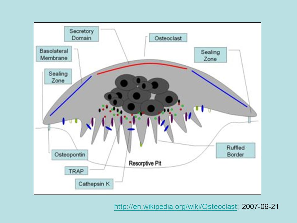
http://en.wikipedia.org/wiki/Osteoclast; 2007-06-21
13
www.drstoute.com/procedures/path1.htm ameloblastoma

14
www.cda-adc.ca/.../graphics/russell_figures.htm cuspid resorption

15
http://www.unige.ch/cyberdocuments/theses2002/MeiriSD/images/image051.jpg ankylose

16
Mavragani et al., 2000

18
Karies?

20
Nordahl, Mjør, Haapasalo, Ørstavik

21
Where are the microbes? AP P PDL

22
Infected dentin? Side connections?
No cells available to start digesting Microtrauma? Etter Haapasalo 2004

23
Surface repair resorption
---- Ankylosis Inflammatory root resorption

24
Behandling av intern resorpsjon
Elisabeth Samuelsen

28
Elisabeth Samuelsen

29
Fuss et al. 2003

31
Heithersay 2004

32
Histologic appearance of an incisor tooth with invasive resorption
Histologic appearance of an incisor tooth with invasive resorption. An intact layer of dentine and predentine on the pulpal aspect (*) separates the pulp from the resorbing tissue. The resorption cavity is filled with a mass of fibrovascular tissue with active mononucleated and multi nucleated classic cells lining resportion lacunae (arrows). (Hematoxylin-eosin stain; original magnification x 40.). (Courtesy of Dr John McNamara.) Heithersay 2004
 separates the pulp from the resorbing tissue. The resorption cavity is filled with a mass of fibrovascular tissue with active mononucleated and multi nucleated classic cells lining resportion lacunae (arrows). (Hematoxylin-eosin stain; original magnification x 40.). (Courtesy of Dr John McNamara.) Heithersay")
33
Heithersay 2004

34
Fig 9 Heithersay 2004

35
Heithersay 2004

36
Histologic appearance of an extensive invasive cervical resorption with radicular extensions. Masses of ectopic calcific tissue are evident both within the fibrovascular tissue occupying the resorption cavity and on resorbed dentin surfaces. In addition communicating channels can be seen connecting with the periodontal ligament (large arrows). Other channels can be seen within the inferior aspect of the radicular dentine (small arrows). (Hematoxylin-eosin stain; original magnification x30.) Heithersay 2004

37
A low powered photograph shows the walling off of the pulp space by dentin separating it from the surrounding extensive resorptive process Fig 12 Heithersay 2004

38
Mass of fibrovascular tissue infiltrated with inflammatory cells, located within a large resorptive cavity that has a wide connection with the periodontal tissue (large arrow). The dentin has been extensively replaced by bone-like tissue. A small section of intact pulp can be seen on the superior aspect of the section (small arrow). Hematoxylin-eosin stain; original magnification x30.) Heithersay 2004

39
Heithersay 2004

40
Heithersay 2004

41
Heithersay 2004

42
Treatment non-surgical treatment involves topical application of a 90% aqueous solution of trichloracetic acid to the resorptive tissue, curettage, endodontic treatment where necessary, and restoration with glass-ionomer cement. Adjunctive orthodontic extrusion may be employed in some advanced lesions. Heithersay 2004

43
Fig 18a Heithersay 2004

47
Invasive Cervical Resorption
Class 1 – Denotes a small invasive resorptive lesion near the cervical area with shallow penetration into dentine. Class 2 – Denotes a well-defined invasive resorptive lesion that has penetrated close to the coronal pulp chamber but shows little or no extension into the radicular dentine. Class 3 – Denotes a deeper invasion of dentine by resorbing tissue, not only involving the coronal dentine but also extending into the coronal third of the root. Class 4 – Denotes a large invasive resorptive process that has extended beyond the coronal third of the root. Heithersay 2004

48
Heithersay 2004

49
Heithersay 2004

51
The resorptive process
Denudation: Cementum Predentin Remodelling: Deposition resorption Infectious/pathological Internal inflammatory External inflammatory Physiological/protective Pressure induced Surface repair Replacement/ankylosis

52
Eksamensspørsmål

53
List opp vilkårene for tannresorpsjon
Dentin må eksponeres: Cementum eller predentin må være brutt Det må være bløtvev med blodtilførsel mot dentin Fra pulpa Fra periodontiet

54
List opp årsaker til tannresorpsjon
Fysiologisk/beskyttende Trykkindusert Overflatereparasjoner Vevsintegrasjon: Ankylose Infeksiøst/patologisk Intern resorpsjon Ekstern inflammatorisk

55
Hva er ”blunting” av røtter? Når skjer det?
Røttene (spesielt overkjevens front) er forkortet og avrundet Forekommer etter aggressiv kjeveortopedisk behandling
 er forkortet og avrundet. Forekommer etter aggressiv kjeveortopedisk behandling.")
56
Klassifisér kliniske former av rotresorpsjon
Lokale resorpsjoner reparerer mikroskader i cement (ikke synlige klinisk el røntgenologisk) Forbigående rotresorpsjon (etter mindre traume) Trykkindusert rotresorpsjon (ortodonti, tannfrembrudd, tumorer) Infeksjonsindusert rotresorpsjon Intern rotresorpsjon Ekstern inflammatorisk rotresorpsjon Erstatningsresorpsjon (ankylose) Cervikale resorpsjoner Isolerte Multiple
 Forbigående rotresorpsjon (etter mindre traume) Trykkindusert rotresorpsjon (ortodonti, tannfrembrudd, tumorer) Infeksjonsindusert rotresorpsjon. Intern rotresorpsjon. Ekstern inflammatorisk rotresorpsjon. Erstatningsresorpsjon (ankylose) Cervikale resorpsjoner. Isolerte. Multiple.")
57
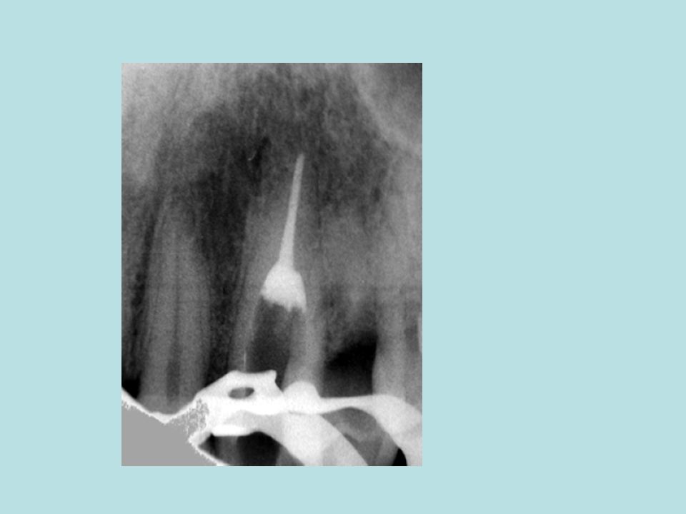
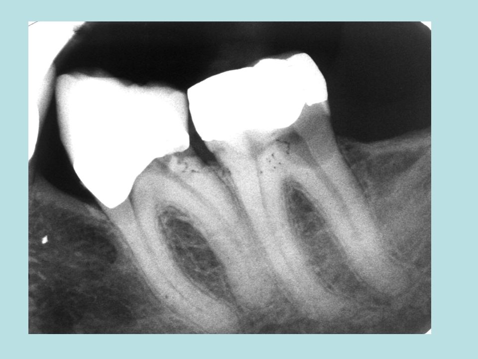
Hva er typisk for intern rotresorpsjon?
Klinisk Gjerne asymptomatisk Uten tegn, Men kan være brutt gjennom og gi symptomer på periodontitt eller (sjelden) frakturere Røntgenologisk Jevn, nær sirkulær Sentrert ut fra pulpa Histologisk Nekrose koronalt, vitalt apikalt (en stund)
 frakturere. Røntgenologisk. Jevn, nær sirkulær. Sentrert ut fra pulpa. Histologisk. Nekrose koronalt, vitalt apikalt (en stund)")
58
Hva er typisk for ekstern inflammatorisk rotresorpsjon?
Klinisk Gjerne asymptomatisk Kan forløpe svært hurtig Følger gjerne et traume (intrusjon, eksartikulasjon) Røntgenologisk Eksentriske opptak vil vise at periodontalspalten er involvert Histologisk Nekrotisk infisert pulpa Ingen spesielle kjennetegn i bløtvevet
 Røntgenologisk. Eksentriske opptak vil vise at periodontalspalten er involvert. Histologisk. Nekrotisk infisert pulpa. Ingen spesielle kjennetegn i bløtvevet.")
59
Hva er typisk for cervikal rotresorpsjon?
Klinisk Gjerne asymptomatisk Pink spot kan forekomme Kan simulere karies I tannhalsen Røntgenologisk ”Møllspist” dentin; ekstensjoner også aksialt i tannen Kan omslutte pulpa; omrisset av den kan gjenkjennes Histologisk Invasjon av osteoid vev i resorpsjonsområdet

60
Beskriv de 4 klassene for cervikal rotresorpsjon
1 Lokalisert i samlet kavitet uten utløpere 2 Starter utbredelse sidelengs og apikalt 3 Begynner å omslutte pulpa, tydelige spor i apikal retning 4 Omslutter pulpa, utløpere apikalt på begge sider av rotkanalen

Liknende presentasjoner


>")


>")


